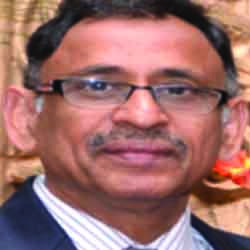
Shalesh Rohatgi
Dr. DY Patil Medical College, IndiaTitle: Study of Clinical, Radiological and Laboratory Features in Neuromyelitis Optica Spectrum Disorder and Myelin Oligodendrocytes Glycoprotein Associated Disorder at A Tertiary Care Hospital in India
Abstract
Abstract:
Introduction-
Inflammatory Demyelinating Disorders (IDD) are emerging as one of the common causes for neurological disability specially in young adults. Neuromyelitis Optica Spectrum Disorders (NMOSD) and anti-Myelin Oligodendrocyte Glycoprotein Associated Disease (MOGAD) are immune-mediated inflammatory conditions of the Central Nervous System (CNS) that frequently involve the optic nerves and the spinal cord. Asians are 3 times and Blacks 10 times more prone to develop NMOSD than Caucasians; thought be linked to certain HLA antigens. Although NMOSD and MOGAD both share a number of common clinical manifestations, they are independent entities with distinct patho-physiological mechanisms and histopathological features. In the present study we studied clinical, radiological and laboratory parameters of the two entities and if there are any differences between the two clinical entities.
Material and Methods-
Type of study- Observational prospective study
Period of study- 3 years from 01 Dec 2020 to 30 Nov 2023
Objective-
1.Primary objective was to study the clinical, radiological and laboratory profile of patients diagnosed with NMOSD or MOGAD
2.Secondary objective was to see if there are any differences in clinical, radiological and laboratory profile between the two groups.
Approval of Institutional ethics committee was taken
Total number of cases-43
Inclusion criteria- Patients who were diagnosed with NMOSD/MOGAD for the first time based on clinical, radiological and laboratory criteria.
Exclusion criteria- Follow up cases of NMOSD /MOGAD were excluded, other etiologies like infectious , metabolic , nutritional , vascular, neoplastic and toxic etiology were ruled out. Multiple sclerosis was excluded.ADEM without MOG- antibodies were also excluded.
Clinical diagnosis of NMOSD was based on the International Panel On NMO Diagnosis (INPD) criteria (2015).
Diagnostic criteria for MOGAD required the presence of a core clinical demyelinating event along with positive MOG- IgG test and exclusion of other diseases.
Results-
43 cases are included, out of which 26 were NMOSD and 17 were MOGAD.
Out 26 patients of NMOSD, 23 were sero-positive and 3 were sero-negative. Radiological investigations included 1.5 TESLA /3 TESLA MRI brain including orbital cuts and spinal cord ( both plain and contrast images). Routine hematological and biochemical parameters, Human immunodeficiency virus, Hepatits-B surface antigen, Veneral Disease Research Laboratory test, Hepatitis C, thyroid profile, anti Aquaporin-4 (NMO) antibodies ( IgG), anti- MOG antibodies, Cerebrospinal fluid ( CSF) analysis, vitamin B12 levels, anti- nuclear antibodies( ANA), anti-neutrophilic-cytoplasmic antibodies, (ANCA) and Visual evoked potentials( VEP) were done in all cases.
Statistical analysis- study is on going .
Statistical analysis using SPSS software will be done after completion of study and shared during presentation
1. Clinical Radiological and Laboratory profile on first presentation- Age ranged from 14 to 70 years. Out of total of 43 patients, majority (44%) were in the third decade , followed by the 4th decade(30%). Mean age at presentation was 33 years .Out of 26 patients of NMOSD 16 (61%) were females , while out of 17 patients of MOGAD 12(70%) were males. Clinical presentation at the onset -unilateral optic neuritis (ON) seen in 6 (23%) patients of NMOSD and 7 (41%) patients of MOGAD . Bilateral ON was seen in3 (11%) cases of NMOSD and 3(17%) cases of MOGAD . Longitudinally extensive transverse myelitis (LETM) was seen in 7 (27%) cases of NMOSD and 4(23%) cases of MOGAD . Short segment myelitis was seen in 1(4%) case of NMOSD and 2 (12%) cases of MOGAD. LETM+ ON was seen in 2(8%) cases of NMOSD and 1(6%) case of MOGAD. Area-postrema syndrome was seen in 5(20%) cases of NMOSD only. Inter-Nuclear Opthalmoplegia (INO) was seen in 2(8%) cases of NMOSD. No cases presented with seizures, encephalopathy, monoplegia or cranial nerve palsies. CSF pleocytosis was seen in 14 (53%) cases of NMOSD and 8 ( 47%) of MOGAD. CSF proteins were raised in 7 (27%) of NMOSD and 8 (47%) of MOGAD. In one patient of MOGAD, CSF oligoclonal bands were also positive.
Assciated autoimmune disease or malignancy- One patient was diagnosed with Sjogren’s syndrome and Hashimoto’s thyroiditis and one patient had non Hodgekins lymphoma in NMOSD.
2. Clinical & Radiological profile on subsequent Relapses-
9(34%) patients of NMOSD and 6 (35%) patients of MOGAD had relapses . Total of 37 relapses occurred in NMOSD group and 15 in MOGAD group . Out of 9 patients of NMOSD who had relapses, 7 were on immunomodulation. In MOGAD group only 1 patient who relapsed was on immuno-modulation.
In relapses; unilateral ON was seen in 12 (32%) relapses of NMOSD and 11 ( 73%) of MOGAD . Bilateral ON occurred in 4 (11%) of NMOSD and 2 ( 13%) of MOGAD relapses.
Spinal cord involvement occurred in 16 (43%)relapses of NMOSD and 2(15%) relapses of MOGAD. Short segment myelitis in 3 (8%) relapses of NMOSD and 2(13%) relapses of MOGAD.Cerebellar ataxia in 1(3%) relapse of NMOSD and none in MOGAD group. Area postrema involvement in 2 (5%) relaspses of NMOSD and none in MOGAD group.
Neuroimaging -
(i)Pattern of involvement of optic nerves -
Out of 16 ( unilateral + bilateral ON) in NMOSD group, LEON was seen in 11(55%), LEON with Chiasmal involvement in 9 (45%) , No intra-orbital or canalicular involvement or perineuitis was seen .
In MOGAD group out of 13 attacks involving optic nerve, LEON was seen in 8( 61%) and chiasmal involvement in 3( 23%). Rest 2 had intraorbital and intra-canalicular involvement. 1 patient had perineuritis.
(ii)Lesions in Brain-
Spratentorial lesions were seen in 6 (35%) relapses of MOGAD and 5 (19%) of NMOSD. Infra-tentorial lesions were seen in 10 (27%) relapses of NMOSD and 4(11%) of MOGAD. In 7(19%) relapses of NMOSD, area postrema involvement was seen .
(iii) Spinal cord-
In 26 relapses of NMOSD who had spinal cord involvement ; LETM was seen in 22(84%). Spinal cord swelling was present in 18 (70%) . Cervical cord involvement was seen in 14 (54%) followed by upper dorsal cord in 6(23%), lower dorsal cord in 2(7%) , cervico- dorsal in 2(7%) and conus medullaris in 1(3%). Extensive cord involvement of entire cord with conus medullaris involvement was seen in 1(3%).
In MOGAD- out of 9 attacks who had spinal cord involvement, 5 were LETM ( 55%). Only in 1 (11%) swelling of spinal cord was present. Cervical and upper dorsal involvement was seen in 3( 33%) , cervical cord only in 2(22%), upper dorsal in 1(11%) and lower dorsal in 1 (11%). Conus medullaris involvement was seen in 2 (22%) of relapses.
Involvement of central and peripheral spinal cord was seen in 14( 54%) in NMOSD, and 3 cases ( 33%) in MOGAD. Only involvement of central cord was seen in 10 relapses (38%) of NMOSD and 5 ( 55%) of MOGAD. Only peripheral involvement of cord was seen in 2 (7%) of NMOSD and 1( 11%) of MOGAD relapses.
Short segment myelitis was seen in 3 ( 8%) of NMOSD and 2(13%) of MOGAD relapses.
Salient findings -
1. NMOSD was more prevalent than MOGAD .
2.Maximum incidence of both NMOSD and MOGAD was in the age group 20-30 years followed by the age group of 30-40 years
3.Female sex predominence was seen in NMOSD and male sex in MOGAD .
4.Unilateral ON was the most common initial presentation and on subsequent relapses in male sex of MOGAD patients.
5.Bilateral ON and LETM were seen equally in both at presentation.
6.Area postrema syndrome occurred only in NMOSD cases.
7. CSF proteins were raised more in MOGAD. Pleocytosis was seen equally in both. One patient of MOGAD also had presence of oligo-clonal bands in CSF.
9.Percentage of patients who had relapses was same in both groups , however total number of relapses were significantly higher in NMOSD group inspite of immunomodulation.
10. Frequency of involvement of spinal cord was almost equal in both the groups at presentation.
11.In subsequent relapses LETM was seen only in NMOSD and none in MOGAD group.
12.Short segment myelitis was almost equal in both groups
13. spinal cord swelling and involvement of both central and peripheral regions of cord was more associated with NMOSD while only central cord involvement was more common in MOGAD
14.Cervical cord was more frequently involved in NMOSD while cervico- dorsal region was more frequently involved in MOGAD
15. Involvement of conus medullaris was seen in both groups equally. Involvement of entire cord with involvement of conus medullaris was seen in one case of NMOSD
16. LEON was seen equally in relapses of both groups. However LEON with chiasmal involvement was seen more in NMOSD. Perineuritis was seen only in one patient of MOGAD
17. Occurrence of supratentorial lesions were more in MOGAD and infratentorial lesions were more in NMOSD.
18. Area postrema syndrome occurred exclusively in NMOSD
19. Occurrence of INO was equal in both the groups.
Conclusion – Despite both NMOSD and MOGAD having many similar clinical and radiological features , there are some differences between them in incidence ,sex distribution, clinical presentation ,relapses, radiological and laboratory findings and response to immune-modulation.
Biography
Surg V Adm( Dr) Shalesh Rohatgi, VSM (Veteran), currently serves as the Professor and Head of the Department of Neurology at Dr DY Patil Medical College, Hospital & Research Centre in Pune. He earned his MBBS from KGMC LKO in 1975, followed by an MD in Medicine from AFMC in 1985, and a DM in Neurology from IMS BHU in 1994. Rohatgi has held various prestigious appointments throughout his career, including Professor and HOD of the Department of Internal Medicine at AFMC, Consultant Neurology for the Armed Forces Medical Services, Honorary Surgeon to the President of India, and Director General Medical Service for the Indian Navy. His contributions to academia are substantial, with 65 papers published in National and International Journals, and authorship of a chapter in the Handbook of Neurological Emergencies. He is a recognized guide and teacher for multiple universities including MUHS, Delhi University, Kurukshetra University, and Dr DY Patil University, as well as a recognized PhD guide at DPU, and serves on the Union Public Service Commission panel of doctors. Rohatgi is also an esteemed member of various neurological societies, including the Indian Academy of Neurology, Indian Stroke Association, and Neurological Society of India. Additionally, he holds associate membership in the International Parkinsons and Movement Disorder Society. His contributions to the field have been acknowledged with honors such as the Vishist Sewa Medal and the Chief of Naval Staff Oration in the Marine Medical Society conference of India in 2013.

